


Project 3: Fetal programming of heart disease in adulthood
Unfavorable intrauterine growth conditions have been shown to predispose the heart for cardiac disease later in life, a process referred to as fetal programming. Generally, intrauterine growth restriction (IUGR) often results in reduced heart size at birth characterized by an incomplete cardiomyocyte complement. We have recently shown in cHccs+/- mice (see project 1) that neonatal cardiac hypoplasia alters postnatal cardiac growth pattern, resulting in transient normalization of organ size until early adulthood by accelerated cardiomyocyte hypertrophy (i.e. increase in cell size). This compensatory hypertrophy cannot be maintained throughout life time, however, such that heart size is reduced again upon ageing. Neonatal hypoplasia also alters cardiac stress response in adulthood, characterized by an overshooting hypertrophic growth and aberrant molecular growth signaling. Inhibition of the latter results in contractile dysfunction in developmentally impaired hearts upon stress. This clearly confirmed that disturbing prenatal cardiac development renders the response of the postnatal heart to pathological conditions. Based on these data we propose a new model linking fetal programming to growth and organ size control in the adult and ageing heart. Surprisingly, adult cHccs+/- mice subjected to left ventricular pressure overload show a better functional outcome compared to controls despite a reduction in cardiomyocyte number. These data suggest that cardiac hypoplasia itself does not seem to negatively affect the response of the adult myocardium towards pressure overload. Instead, it raises the interesting hypothesis that altered intrauterine cardiac growth could condition the heart for certain pathological stimuli during postnatal life.
We are currently investigating the consequences of different IUGR models for growth of the pre- and postnatal heart as well as cardiac stress response in adulthood. A special focus lies on mTOR (mechanistic target of rapamycin), an important regulator of translation and cell growth. Whereas mTOR has been intensively studied in the context of pathological as well as physiological cardiac hypertrophy in adulthood, surprisingly little is known about its relevance for the prenatal heart. Therefore, we developed a rapamycin treatment protocol in pregnant mice to efficiently induce IUGR in the offspring and investigate the importance of mTOR for perinatal cardiac growth. Current projects intent to further study its role during embryonic and fetal heart development using genetic models of heart conditional inactivation of mTOR components in mice.
IUGR in humans is often caused by impaired nutrient supply to the embryo or fetus, which can be a result of maternal undernutrition or placental insufficiency (among others). In animal models IUGR can be induced by protein restriction in the maternal diet during pregnancy, which in the offspring results in cardiac hypoplasia at birth due to a reduced number of cardiomyocytes. To assess the importance of amino acid availability for cardiac growth and organ size control, we are investigating the consequences of a low protein diet throughout pregnancy as well as during postnatal life in mice and rats. Our preliminary results suggest that amino acid restriction does not alter developmental patterning or growth of the embryonic heart, but results in reduced heart size in adult mice if kept on a low protein diet after birth. Thus, sufficient amino acid availability appears to be primarily important for postnatal hypertrophic but not prenatal proliferative growth of the heart. Current projects intend to thoroughly characterize the role of amino acid availability during the transition from pre- to postnatal cardiac growth pattern as well as a possible influence on myocardial cell composition and tissue homeostasis after birth.
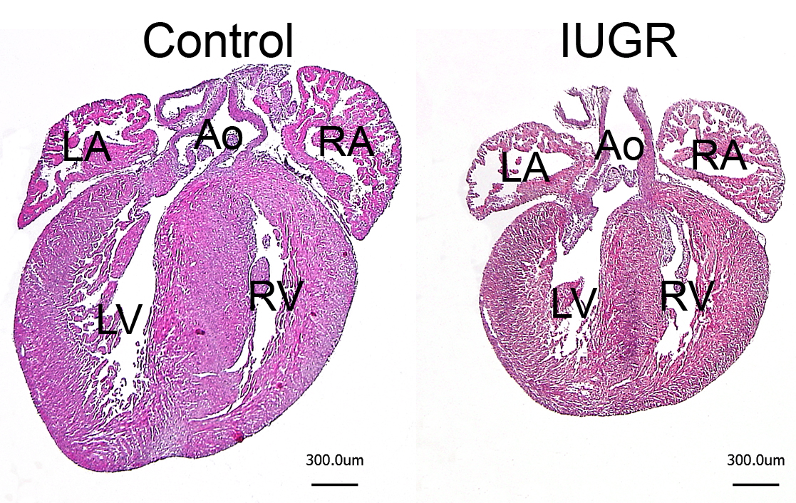
Figure 3: Intrauterine growth restriction (IUGR) reduces heart size at birth.
IUGR was induced by mTOR (mechanistic target of rapamycin) inhibition during late gestation in mice. Newborn hearts of the resulting offspring were embedded in paraffin, sectioned and stained with hematoxylin & eosin (H&E). Overview images show a striking reduction in heart size in IUGR compared to control pups. LV/RV = left and right ventricle, LA/RA = left and right atrium, Ao = aorta (scale bar = 300 μm)
Related Publications:
Drenckhahn JD, Strasen J, Heinecke K, Langner P, Yin KV, Skole F, Hennig M, Spallek B, Fischer R, Blaschke F, Heuser A, Cox TC, Black MJ, Thierfelder L. Impaired myocardial development resulting in neonatal cardiac hypoplasia alters postnatal growth and stress response in the heart. Cardiovasc Res 2015;106:43-54.
doi: 10.1093/cvr/cvv028.
Heinecke K, Heuser A, Blaschke F, Jux C, Thierfelder L, Drenckhahn JD. Preserved heart function after left ventricular pressure overload in adult mice subjected to neonatal cardiac hypoplasia. J Dev Orig Health Dis 2017 Jul 24:1-13.
doi: 10.1017/S2040174417000514.
Hennig M, Fiedler S, Jux C, Thierfelder L, Drenckhahn JD. Prenatal Mechanistic Target of Rapamycin Complex 1 (mTORC1) Inhibition by Rapamycin Treatment of Pregnant Mice Causes Intrauterine Growth Restriction and Alters Postnatal Cardiac Growth, Morphology, and Function. J Am Heart Assoc 2017;6(8). pii: e005506.
doi: 10.1161/JAHA.117.005506.
Links:
https://goo.gl/5o2AFn
https://goo.gl/j3t7Mu
https://goo.gl/m1ywmb